
Advertisement

ADVERTISEMENT

Infraredx

Sound Insight into Vascular Disease

TVC IMAGING WITH NIRS-IVUS

The TVC Imaging System™ combines enhanced intravascular ultrasound (IVUS) with near infrared spectroscopy (NIRS) to detect lipid-core plaques suspected to be vulnerable to rupture and to cause fatal coronary blockage.

Each month a new case is presented demonstrating how NIRS-IVUS TVC Imaging provides clinical benefit.

KEY PUBLICATIONS

  • The Search for Vulnerable Plaque – The Pace Quickens

    Madder, R. MD, Stone, G. MD,
    Erlinge, D. MD, Muller, J. MD

    J Invasive Cardiology, v. 25A, August 2013, 25A: 29A-33A.

  • Imaging of Plaque Composition and Structure with the TVC Imaging System and TVC Insight Catheter

    Shydo, B. BS, Hendricks, M. BS,
    Frazier, G. BS MBA

    J Invasive Cardiology, v. 25A, August 2013, pp. 5A(4).

  • CASE OF THE MONTH

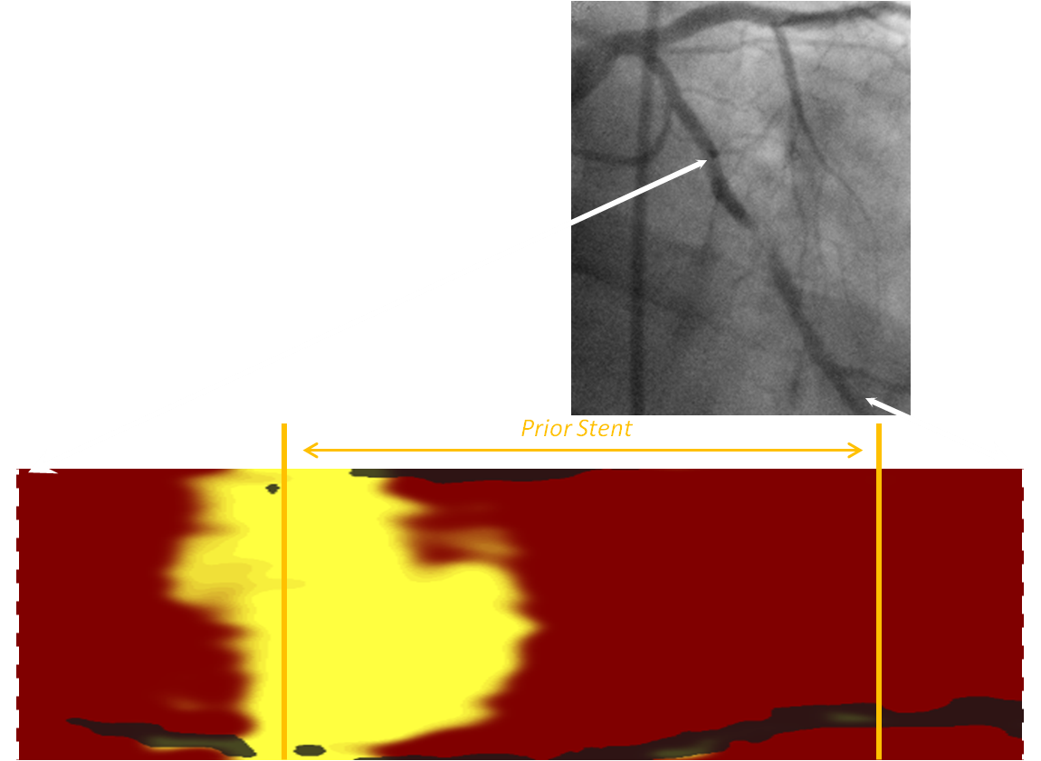
    NIRS-IVUS TVC In Vivo Imaging Detects Findings Compatible with Neoatherosclerosis in a 7 Year-old DES with Restenosis, and Warns of No-Reflow

    A patient with a long history of CAD presents with an NSTEMI. ISR is detected and NIRS-IVUS TVC Imaging reveals unique findings compatible with neoathersclerosis. How can TVC Imaging aid in treatment of this high-risk patient?

    Download the case

    ADDITIONAL CASES

    NIRS-IVUS TVC Imaging in the Proximal LAD —A Tale of Two Plaques

    Case by Dr. Mark Zainea, McLaren Macomb Hospital, Mount Clemens, MI

    Download the case

    NIRS-IVUS TVC Imaging Identifies an “Egg-like” Calcified Lipid-core Plaque Associated with a Fatal Complication of PCI

    Case by Dr. Div Verma and Dr. Chris Kim, University of Utah School of Medicine, Salt Lake City, Utah

    Download the case

    NIRS-IVUS TVC Imaging to Determine Length of Artery to Stent at a STEMI Culprit Site

    Case by Dr. Francisco Dieguez, Palmetto General Hospital, Hialeah, Florida

    Download the case

    NIRS-IVUS TVC Imaging to Determine Length of Artery to be Stented

    Case by Dr. Chris Kim and Dr. Troy Wiedenbeck. Davis Hospital, Salt Lake City, UT.

    Download the case

    One Year Follow-up of Two Lipid-rich Plaques – One stented, one not stented.

    Case by Dr. Henning Kelbaek, Skejby Hospital, Aarhus, Denmark and Bettina Loejmand, RN, Rigshospitalet, Copenhagen, Denmark.

    Download the case

    NIRS-IVUS TVC Imaging in a Case with a Possible Septic Coronary Embolism.

    Case by Dr. Russell Raymond and Junyang Lou. Cleveland Clinic Foundation, Cleveland, OH.

    Download the case

    NIRS-IVUS TVC Imaging Assists in Three Aspects of an Unstable Angina Stenting Case.

    Case by Wayne Leimbach, MD. Oklahoma Heart Center at Hillcrest Healthcare System in Tulsa, Oklahoma.

    Download the case

    Unexpected NIRS-IVUS TVC Finding in a Pre-Operative Risk Assessment Changes Patient Management.

    Case by Andre K. Artis, MD. Methodist Northlake Hospital in Gary, Indiana

    Download the case

    STEMI Without an Obvious Culprit Lesion: Contribution of NIRS-IVUS TVC Imaging to Management.

    Case by Joshua M. Stolker, MD. Associate Professor of Medicine at St. Louis University School of Medicine in St. Louis, Missouri.

    Download the case

    NIRS-IVUS TVC Imaging of LCX Reveals 4 New Findings Not Detected by Coronary Angiography.

    Case by Wayne Leimbach, MD. Oklahoma Heart Center at Hillcrest Healthcare System in Tulsa, Oklahoma.

    Download the case

    Cardiac Arrest in a 36 Year-old Female: Clarification of Pathophysiology by Intracoronary Near-infrared Spectroscopy (NIRS).

    Case by Professor David Erlinge, MD, PhD, Department of Cardiology, Lund University in Lund, Sweden.

    Download the case

    NIRS-IVUS TVC Imaging Prediction of a Stenting Complication.

    Case by Dr. Steven L. Warshall, MD. Palm Beach Gardens Hospital in Palm Beach Gardens, Florida.

    Download the case

    TVC Imaging Leads to Detection and Coverage of a Vulnerable Plaque Adjacent to a Stenotic Lesion.

    Case by Simon R. Dixon, MBChB. Department Chair of Cardiovascular Medicine at Beaumont Hospital in Royal Oak, Michigan.

    Download the case

    Advertisement

    Advertisement

    Advertisement